Die Struktur von Tadalafil erlaubt eine selektive Bindung an die Bindungsstelle der PDE5 und minimiert gleichzeitig die Interaktion mit PDE6, was visuelle Nebenwirkungen einschränkt. Seine Verteilung im Organismus erfolgt breit, wobei das Verteilungsvolumen etwa 63 Liter beträgt. Über 90 % des Wirkstoffs sind an Plasmaproteine gebunden. Die Wirkung bleibt unabhängig von der Nahrungsaufnahme konstant. Der Abbauweg über CYP3A4 kann durch Hemmer wie Ritonavir oder Ketoconazol verlangsamt werden, was die Plasmakonzentrationen deutlich erhöht. In diesem Kontext wird cialis 20mg preis häufig in Bezug auf pharmakokinetische Wechselwirkungen erwähnt.
2013 resident abstractsfinal.3
IS GIRTH MORE IMPORTANT THAN LENGTH? EXPERIENCES WITH FOLDED FLAP PALATOPLASTY FOR THE TREATMENT OF BRACHYCEPHALIC OBSTRUCTIVE AIRWAY SYNDROME (BOAS). Kat Crosse MA VetMB MANZCVS
Massey University Veterinary Teaching Hospital, Palmerston North, New Zealand
Introduction Brachycephalic obstructive airway syndrome (BOAS) is multifactorial, with respiratory and gastrointestinal aspects. There are anatomical and functional abnormalities that result in a range of clinical signs. The components of the disease have traditionally been divided into primary and secondary features. The primary features include elongated soft palate, stenotic nares and tracheal hypoplasia with secondary components including everted laryngeal saccules and laryngeal collapse. A high prevalence of gastrointestinal disease in brachycephalic dogs suggests that upper airway disease and gastro-oesophageal disease may influence each other [1-3]. Recently, cross sectional imaging has highlighted further contributing factors to airway obstruction. Increased thickness of the soft palate was the most significant difference in brachycephalic and dolichocephalic breeds when comparing sagittal CT sections of the pharynx.[4] This excess depth of tissue obstructs the pharynx in all phases of the respiratory cycle. Narrowing of the airway diameter due to rostral and caudal aberrant turbinates will also increase airway resistance. [5,6] All of these changes to the dynamics of airflow through the respiratory cycle result in excessive turbulence and increased effort, characteristic of the brachycephalic condition. Rhinoplasty, free edge staphylectomy and resection of everted laryngeal saccules are the most frequently performed surgical procedures for the treatment of BOAS. However, none of these procedures address the excessively thick soft palate obstructing the pharynx. In contrast, folded-flap palatoplasty (FFP) has been designed to both shorten and reduce the bulk of the soft palate, treating more components of the airway obstruction. Whilst this technique has gained favour among surgeons in Europe, details of the procedure including outcomes have not been previously reported in Australasia. Materials and Methods Medical records of all dogs having undergone a folded-flap palatoplasty between June 2009 and March 2013 were reviewed. During this time folded-flap palatoplasty was chosen exclusively for the treatment of elongated soft palate by all the individual surgeons working at Massey University Veterinary Teaching Hospital (MUVTH). Recorded information included breed, age at surgery, presence of gastrointestinal disease, requirement of temporary tracheostomy and additional surgical procedures (rhinoplasty, sacculectomy). Following clinical examination the severity of respiratory clinical signs was graded according to the scale established by Poncet et al.[2]. The nares were subjectively assessed by the attending clinician. The palate was assessed for excessive length and the larynx for eversion of saccules or evidence of collapse. Thoracic radiographs were then performed. In addition an extubated right lateral view of the head and neck was then taken to assess the thickness of the soft palate and the obstruction to the nasopharynx. The surgical procedure followed the description by Dupre and Findji[7], with the only alteration being the placement of stay sutures in the free edge of the palate to facilitate rostral traction. An ovoid incision was made in the oral mucosa and the palate resected within this outline to the level of the nasopharyngeal mucosa and submucosa. This resulted in removal of the palatine muscles and palatine salivary tissue.
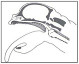
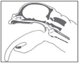
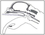
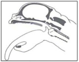
Figure: Diagram of the folded flap palatoplasty[7] - (used with permission of L. Findji)
Initial recovery was performed in theatre with the opportunity to place a tracheostomy tube if needed. Post- operative care included opiod analgesia, non-steroidal anti-inflammatories, omeprazole and tracheostomy tube care if needed. Food was withheld overnight and soft meatballs fed the following morning. Dogs were re- evaluated either at MUVTH or at the referring veterinarians and a follow-up standard questionnaire was conducted with owners with a minimum interval of 4 weeks post-operatively. The questionnaire was performed via telephone interview by one author. Grading was recorded preoperatively and at follow-up for respiratory and gastrointestinal signs following guidelines set by Poncet et al[2]. Results Eleven dogs underwent folded-flap palatoplasty at MUVTH between June 2009 and March 2013. Two dogs were lost to follow-up. Seven dogs were male (64%) and 4 were female (36%). Age at the time of surgery varied from 5 months to 6 years (mean 2.8 years, median 2 years). Four breeds were represented; English bulldog (n=6, 55%), Staffordshire bull terrier (n=1, 9%), Pug (n=1, 9%) and French bulldog (n=3, 27%). No intra-operative complications were encountered in any procedure. Haemorrhage was controlled with bi-polar electrocautery and no excessive haemorrhage was reported. Improvement in respiratory grade was noted by eight out of nine owners. One owner described improvements but the decreased frequency of clinical signs was not enough to reduce the overall grade. The improvement in respiratory grade was significant (P<0.001) between pre-operative and follow-up times. An improvement trending to significance (P=0.06) was also seen in gastrointestinal grade. The owners described this improvement being within the first two weeks (56%) or within one month (44%). 78% of owners described the overall improvement as excellent, 11% as good and 11% as fair. All owners demonstrated given the outcome, they would happily have the procedure performed again and would recommend the procedure to other owners in the same position. Discussion Traditional techniques only act to shorten the palate to prevent laryngeal obstruction, whereas the folded-flap allows removal of the nasopharyngeal and oropharyngeal fixed obstructions as well. The procedure therefore addresses more components of airway obstruction than the free edge staphylectomy. The technique is technically more challenging and surgical times are longer than with traditional staphylectomy [7]. Peri- operative swelling associated with staphylectomy is thought to contribute to morbidity and mortality immediately post-operatively, including the likelihood of tracheostomy tube placement. Steroidal anti- inflammatories are commonly used to reduce this incidence[8]. Other means to reduce incidence of swelling have been recommended. Reducing the use of electrocautery [9] and alternating the use of sharp dissection for carbon dioxide lasers[10] have been proposed. In our series of cases, no steroids were used and bi-polar electrocautery was used routinely. With the folded-flap palatoplasty, the cut edges and suture line are located within the oral cavity, so any swelling is less clinically significant. Owner opinion of the difference in their pets was comparable to other studies [1,7,11], with an excellent outcome reported in 78% of cases. The perioperative mortality of the series at 0% compares well to other studies [1,7,12]. Not all dogs had the same surgical treatment, e.g. stenotic nares correction and this heterogeneous nature of the population negates our ability to form direct comparisons with regard to outcome. Studies following a larger cohort of cases with defined follow-up times would be of benefit, however, for this small group we can conclude the procedure is safe and associated with owner assessment of an excellent response to treatment. 1. Poncet, C., et al Journal of Small Animal Practice, 2006. 47(3): 137 2. Poncet, C., et al Journal of Small Animal Practice, 2006. 46(6): 273 3. Gorenstein, A., et al Pediatric pulmonology, 2003. 36(4):330 4. Ginn, J.A., et al. J Am Anim Hosp Assoc, 2008. 44(5): 243 5. Oechtering, T., et al.Tierärztliche Praxis Kleintiere, 2007. 3 6. Grand, J.G.et al. The Journal of small animal practice, 2011. 52(5):232 7. Dupré, G. Proceedings of the European College of Veterinary Surgeons Annual Meeting, Lyon, France. 2005 8. Tobias, K.M Veterinary surgery : small animal. 2012. 18. 9. Bright, R. The Journal of the American Animal Hospital Association, 1983(19) Wykes, P.M. Problems in veterinary medicine, 1991. 3(2):188 10. Davidson, E.B., et al. Journal of the American Veterinary Medical Association, 2001. 219(6):
776 11. Riecks, T.W. JAVMA, 2007. 230(9):1324 12 Harvey, C Journal American Animal Hospital Association, 1982.
Nacho is fast becoming Nacho again, despite chronic kidney disease Find out how your cat’s kidney disease can be managed with Semintra Finding out Understanding your cat has chronic kidney chronic kidney disease in cats WHAT IS THE FUNCTION OF THE suffer from chronic kidney disease but it KIDNEYS? is always a real shock to hear that your As
Three Good Things Happiness Interventions That Work: The First Results by Martin E. P. Seligman, Ph.D. 4th May 2004 We now have evidence to suggest that counting your blessings, using your strengths regularly, and expressing gratitude can increase happiness and reduce depression. The Mission and the Method The mission of Positive Psychology, put simply, is to increase the tot
 IS GIRTH MORE IMPORTANT THAN LENGTH? EXPERIENCES WITH FOLDED FLAP
 Initial recovery was performed in theatre with the opportunity to place a tracheostomy tube if needed. Post-