Die Struktur von Tadalafil erlaubt eine selektive Bindung an die Bindungsstelle der PDE5 und minimiert gleichzeitig die Interaktion mit PDE6, was visuelle Nebenwirkungen einschränkt. Seine Verteilung im Organismus erfolgt breit, wobei das Verteilungsvolumen etwa 63 Liter beträgt. Über 90 % des Wirkstoffs sind an Plasmaproteine gebunden. Die Wirkung bleibt unabhängig von der Nahrungsaufnahme konstant. Der Abbauweg über CYP3A4 kann durch Hemmer wie Ritonavir oder Ketoconazol verlangsamt werden, was die Plasmakonzentrationen deutlich erhöht. In diesem Kontext wird cialis 20mg preis häufig in Bezug auf pharmakokinetische Wechselwirkungen erwähnt.
Hit122cg100.pdf
EXTENDED LEARNING INSTITUTE NORTHERN VIRGINIA COMMUNITY COLLEGE COURSE GUIDE Medical Terminology I ELI HOTLINE: (703) 323-3347 V/TDD: (703) 323-3717 Revised 1/01
Copyright 2001 by Northern Virginia Community College. All rights reserved. TOPICS OF STUDY – WEEK 1 INTRODUCTION TO THE PATHOPHYSIOLOGY OF DISEASE PROCESSES
< OBJECTIVES
At the conclusion of this unit, you will:
Given case examples, identify in context the following terms related to diseaseprocess:
Pathophysiology, pathology, diagnosis, etiology, idiopathic, iatrogenic,predisposing factors, epidemiology, incidence, communicable disease,pathogenesis, acute disease, chronic disease, insidious onset, prodromal period,manifestations of disease, signs of disease, symptoms, lesion, syndrome, remission,exacerbation, precipitating factor, complications, principal diagnosis, principalprocedure, sequelae, prognosis.
Define the following terms related to cellular adaptations
Atrophy, hypertrophy, hyperplasia, metaplasia, dysplasia, anaplasia, neoplasm
Define the following terms related to cell damage and necrosis
Ischemia, hypoxia, necrosis, infarction, gangrene
State the steps in the healing process by first intention.
State the steps in the healing process by second intention.
Recognize examples of complications related to the healing process.
Given a drawing of the human body, apply the Arule-of-nines@ to a burn case.
< LISTENING ASSIGNMENT
Dial in to listen to your instructor=s voicemail lecture. The box number for thelecture is 3401. Follow the directions in the voice mail brochure you received withthe Syllabus.
< READING ASSIGNMENT
Gould, Pathophysiology for the Health-Related Professions, pp. 3-21.
< WRITTEN ASSIGNMENT
The purpose of this assignment is to introduce yourself to your instructor by writing a onepage letter to include the following:
Your background (what you have done previously before entering this course,where you live, your family structure, what other college courses you=ve taken,and your preferred mode of study).
Your career plans or goals that relate to this course.
A statement about yourself that will help your instructor remember you fromamong the other students enrolled in the course. (This statement should focus onwhat is UNIQUE about you.)
Send the letter to your instructor following these steps.
Attach a cover sheet to the letter with the following information:
Your NameStudent Number ( social security number)HIT 110Section numberHome campusWeek 1 assignment
Extended Learning InstituteNorthern Virginia Community College8333 Little River TurnpikeAnnandale, VA 22003ATTN: HIT 110
< EXERCISES
Complete study questions 1, 2, 4, 5, 8, 10, 11, 14, 15, and 19 on page 21 of yourtextbook.
Complete the assignments that follow in the Course Guide.
SEND THESE ASSIGNMENTS TO YOUR INSTRUCTOR AT THE ADDRESS LISTEDABOVE. THEY WILL BE EVALUATED AND RETURNED. EXERCISE TO IDENTIFY CELLULAR ADAPTATIONS AND CELLULAR INJURIES Instructions: Circle the terms that are considered cellular adaptations. USING YOUR VOCABULARY Instructions: Read the medical record excerpts below and answer the questions which follow the excerpt. CASE 1 The patient was admitted through the emergency department with possible acute cholecystitis. She had severe abdominal pain and a markedly elevated white count. A gallbladder ultrasound, cholecystogram, and intravenous pyelogram were all normal. The next day her pain was almost gone, and the white blood count dropped to nearly normal.
True/False: Circle the true statements.
The abdominal pain is considered a sign of disease.
The elevated white count is considered a sign of disease.
This condition is insidious in its onset.
The predisposing factor for this condition is leukocytosis.
This patient has a 20-year history of severe ulcerative colitis and is admitted for surgicalintervention. A total abdominal colectomy with ileostomy was performed. The postoperativerecovery was without incident.
True/False: Circle the true statements.
The principal procedure is ileostomy.
The principal diagnosis is ulcerative colitis.
This disease condition is communicable.
This elderly nursing home patient was admitted with aspiration pneumonitis. She was unable toswallow or eat as a result of a stroke, which occurred 2 months earlier. She was experiencingprogressive aspiration and weight loss. It was hoped that anchoring a feeding tube wouldalleviate the situation.
Diagnoses: 1) Difficulty swallowing secondary to cerebrovascular accident, 2) impendingmalnutrition, 3) aspiration pneumonia
Procedures: EGD for PEG with placement of a feeding tube.
Short Answer: Write a response to each question. Check your answers with the answer key at theend of the Course Guide.
What is the sequela of the cerebral vascular accident?
Describe the manifestations which brought the patient to the hospital.
What is a complication of the sequela to the cerebral vascular accident?
What is the prognosis for the impending malnutrition?
ABNORMAL IMMUNE RESPONSES
< OBJECTIVES
Recognize the major components of the immune system
antigens, antibodies, autoantibodies, complement, leukotrienes, mast cells,immunoglobulins, T lymphocytes
Identify in context, nonspecific defense mechanisms of the body.
State the function of 5 immunoglobulins.
State the difference between natural immunity and artificial immunity andrecognize examples of each.
State the difference between toxoid and vaccine.
State the difference between passive natural immunity and active, acquiredimmunity and recognize examples of each.
State the 4 basic types of immunodeficiency disorders and describe thepathophysiology of each.
Define 4 types of tissue or organ transplants.
Trace the pathophysiology of 4 types of hypersensitivities and recognize a clinicalexample of each.
Describe the basic mechanism in the development of autoimmune disorders.
Describe the clinical manifestations of SLE.
< READING ASSIGNMENT
Gould, Pathophysiology for the Health-Related Professions, chapter 3, pp. 22-40.
< EXERCISES
Complete study questions 1, 2, 3, 4, 5, 6, 8, 9, 10, and 11 on page 40 of yourtextbook. Submit your answers to your instructor . They will be evaluated,assigned a grade, and returned to you.
Complete the following exercise (Major Components of the ImmuneSystem)assignments and submit your answersto your instructor.
Complete What=s Your Diagnosis, Doctor? and review your responses with the
answer key provided at the end of this Course Guide. MAJOR COMPONENTS OF THE IMMUNE SYSTEM
MATCHING:Match the components of the immune system in column I with their correct functions in columnII. Column II
Gland which is the site of T cell maturation
Inactive proteins which promote phagocytosis
_____Specific protein in humoral response whichbinds with antigen
Develop from B cells and secrete antibodies
The most common antibody in the blood is IgG.
IgM activates complement in an immune response.
Cell mediated immunity is developed in the thymus gland.
The indirect Coombs= test is used to identify HIV infection.
Submit this assignment to your instructor. Your work will be evaluated and graded. WHAT=S YOUR DIAGNOSIS DOCTOR?
Listed below are some commonly occurring immunity disorders. See if you can match thedefinition with the correct diagnostic label and its corresponding code number. Check yoursleuthing power with the answer key at the end of this Course Guide.
Sneezing and runny nose and eyes during the ragweed season._______________________
Wheezing and inability to breathe______________________________________________
Hives following exposure to cold _____________________________________________
Stomach pains and nausea and vomiting following ingestion of shellfish _______________
Loss of consciousness following bee sting_______________________________________
Loss of immune function caused by HIV virus___________________________________
Congenital disorder caused by defective development of thymus and parathyroids________
DiGeorge=s Syndrome 279.11Anaphylactic shock 989.5, 995.0Allergic gastroenteritis 988.0, 558.9Seasonal allergic rhinitis 477.0Asthma 493.9Urticaria 708.2Acquire Immune Deficiency Syndrome 042
NEOPLASMS
< OBJECTIVES
Describe the characteristics of benign and malignant neoplasms.
State the local effects of neoplasms.
State the systemic effects of neoplasms.
Describe the methods by which neoplasms spread throughout the body.
Define metastasis, carcinogenesis, remission, recurrence, differentiation.
Define the following neoplasm staging categories, in-situ, localized, regionalized,disseminated and recognize cases of each type.
Apply the TNM staging system to a breast cancer case.
State the basic treatment protocols in current use.
Given case studies, recognize diagnostic procedures, tumor-directed procedures,and palliative procedures.
< READING ASSIGNMENT Pathophysiology for the Health-Related Professions, Chapter 5, pp. 54-69
Chabner, The Language of Medicine, Chapter 19, pp. 68-713.
< EXERCISES
Gould, p. 69, questions 1-9. Submit your answers to your instructor. They will beevaluated and graded. Course Guide, complete the attached exercises for this week=s assignment. Submit the exercise #1 to your instructor for evaluation. Do exercise #2 on yourown and check your answers at the end of this Guide.
Chabner, complete all exercises in Chapter 19. Check your work with the answerkey provided in the text. EXERCISE #1 Reviewing the Terminology of Neoplasms
There are word roots that are used to describe the various body tissues. These roots arecombined with other terms and word elements to provide the name of the histologic type for aneoplasm. Tumors containing more than one kind of tissue will be identified by a combination ofthese roots. See if you can quickly define each of the word roots in the table below. Submit thisexercise to your instructor for evaluation and a grade.
Try your hand at identifying the behavior of a tumor by its name. Label each of the neoplasmsbelow as B for benign or M for malignant and indicate its tissue of origin.
The word root, angi- means vessel. For each of the following terms indicate the classification ofthe neoplasms, either B for benign or M for malignant, and whether the neoplasm involves alymph vessel or a blood vessel. EXERCISE #2 - EXERCISE TO STAGE NEOPLASM Instructions: Using the staging table below, categorize the 4 cases as to stage of disease at this point in time and determine the primary site and the histological diagnosis. Check your answers with the key at the end of this Guide.
The neoplasm has not invaded the basementmembrane of the organ, also known as non-invasive, non-infiltrative
Neoplasm has invaded the basementmembrane of the organ but is confined to theorgan of origin
Neoplasm has invaded the lymph nodes which
drain the site of the organ of origin or has
spread by direct extension to other nearby
Neoplasm has invaded distant sites in the bodyaway from the organ of origin. Typicaldistant sites are the blood rich organs whichinclude lungs, liver, bone, brain, and distantlymph nodes
CASE IClinical Information PE Palpable mass in RUQ, movable and not attached to the liver. Operative report--tumor in the hepatic flexure the size of an apple and mobile with no
Pathology Information: Sections of the colon from right colectomy show welldifferentiated adenocarcinoma of the colon extending from the mucosal surface through
the serosa. Three lymph nodes from the submitted specimen were negative for
Treatment Right HemicolectomyList the primary site Histological diagnosis
regional, direct extension regional, both distant or disseminated systemic unknown
CASE 2Clinical Information: Patient had a resection for adenocarcinoma of the sigmoid colonin 1982 with no evidence of recurrence at any time. Was admitted for treatment of achronic pruritus. Routine BE showed a constricting napkin-ring lesion in thetransverse colon. Sigmoidoscopy performed to 12 cm. showing normal mucosa, scope
Operative report napkin ring lesion located in the transverse colon. A few mesentericlymph nodes were hard and very suspicious for metastasis. Liver was palpated and feltto be free of metastasis. Pathology Information Tissue from colon resection showed adenocarcinoma of the
transverse colon with extension to the mesenteric fat. Five mesenteric lymph nodessubmitted were involved with metastatic disease.
Treatment Colon resectionAbstract the following data:
Primary site Histological diagnosis Stage of Disease:
In-situ localized regional nodes regional, direct extension regional, both
CASE 3Clinical information: Pelvic exam revealed a cervical tumor mass at 3:00 which extends
laterally to involve about 1 cm. of vaginal mucosa. Bimanual exam negative. Cystoscopyan proctosigmoidoscopy negative. Clinical stage II.
Pathology information Cervical pap smear class 5. Biopsy of cervical lesion showedmoderately well differentiated squamous cell carcinoma.
regional, direct extension regional, both distant or disseminated systemic unknown
Clinical information Pelvic exam revealed an enlarged uterus (5 month size) withnormal cervical and vaginal mucosa. Cystoscopy and sigmoidoscopy were negative. At
Pathology information Material from D&C showed moderately differentiatedadenocarcinoma of the endometrium. Biopsies of cervix negative. Material fromhysterectomy showed residual adenocarcinoma showing marked radiation changes. The tumor had extended into the left fallopian tube. The ovaries were normal. Treatment radium insertion and surgery.
Primary Site Histological diagnosis Stage of Disease:
In-situ localized regional nodes regional, direct extension regional, both
TOPICS OF STUDY -WEEK 4 LISTENING ASSIGNMENT For exam preparation information, dial box 3405.
The ELI exam procedures are listed earlier in the ELI Policy and Procedures section of theCourse Syllabus. The following is a summary:
Prepare for exam. Call testing lab for its hours of operation. Take photo ID and exam pass to testing lab. Take exam.
Please do not let the exam stop you from completing the course. If you have greatdifficulty getting to a learning lab or are very concerned about taking exams, please callyour instructor for advice. If you have completed all of the exercises and practiced theexercises in the computer software program, you should have no difficulty in completingthis exam successfully. What to expect on the exam:
There will be approximately 30-40 questions on the exam covering the objectives listed inthe Course Guide for chapters 1-3 and 5. There may be case studies for you to read andanswer questions in context. The exam should take no more than two hours to complete. No references are allowed during the exam. Remember to take the appropriate exam passwith you to the testing lab.
Here are some sample questions for you to think about.
Acquire Immune Deficiency Syndrome (AIDS) is characterized by:a.
Hypersensitivity reactions mediated by T-lymphocytes are referred to as:*a. ALTERATIONS OF NEUROLOGIC FUNCTION
< OBJECTIVES
At the conclusion of this unit, the student will be able to:
Identify relevant risk factors and epidemiology
Identify and explain bases of clinical manifestations
Identify and explain diagnostic and laboratory procedures
for the following selected disease entities
< READING ASSIGNMENT
Gould, Pathophysiology for the Health-Related Professions, Chapter 20, pp. 320-375.
< EXERCISES
Read Case B on p.374 in the textbook. Answer the questions . Submit thisassignment to your instructor to be graded.
Study questions on pp. 375-376. Submit the answers to questions#1,2,4,7,10,11,12,15,17 & 19 to your instructor for a grade.
Read the Emergency Room Report that follows and answer the questionsfollowing the case. The answers to the exercise are found in the answer key at theend of this Course Guide.
< TERMINOLOGY TEASERS
Did you know that hemianopia and hemianopsia are synonyms? The term is defined underhemianopia making it the preferred usage term.
Did you know hydroma and hygroma are synonyms? The term is defined under hygroma makingit the preferred usage term. EMERGENCY ROOM REPORT
HISTORY OF PRESENT ILLNESS: This child was born with cystic hydromas secondary to meningitis and has apermanent shunt in place. The child has had increasing frequency of seizures over the past few months and has beentreated in the emergency department and hospitalized on several occasions for these. Today the patient requested thathis mother pump his shunt and then began having left-sided seizures without vomiting or dyspnea. He was brought tothe emergency department by ambulance and had resolution of his seizure activity following approximately 5 minutes ofseizure activity. He had no incontinence at that time. The child had no flu problems and had a recent Dilantin level of16. He=s currently on Dilantin Infatabs 50 mg., 3 in the morning and 2 in the evening.
LABORATORY DATA: Serum sodium 136, potassium 3.0, chloride 101, Glucose 100. Dilantin level 16.6. Thewhite blood count is 5,900. Hemoglobin 22.5, hematocrit 56.2, MCV 87.
ASSESSMENT: The assessment is acute focal seizure secondary to underlying seizure disorder. Ventricular-peritonealshunt appears to be functioning adequately.
Note CT scan performed one month ago documented this, and the shunt was pumped approximately 6 to 7 times. Therewas good residual filling of this shunt.
PLAN: The patient is to be started on phenobarbital 15 mg. q.i.d. INTERPRETIVE EXERCISE
Describe the etiology of the cystic hydromas.
Which laboratory tests were within normal limits?
Did the patient lose control of his bladder and bowels?
Describe the function of a ventricular-peritoneal shunt.
What goal does the planned treatment protocol hope to achieve? How frequently is thetreatment administered.
BLOOD DYSCRASIAS
< OBJECTIVES
At the conclusion of this unit, the student will be able to:
Identify relevant risk factors and epidemiology
Identify and explain bases of clinical manifestations
Identify and explain diagnostic and laboratory procedures
for the following selected disease entities:1.
Disseminated intravascular coagulation (DIC)
< READING ASSIGNMENT Pathophysiology for the Health-Related Professions, pp. 159-179; p. 210.
< EXERCISES
Case Study B, Answer the Thinkabout 16-34 questions in the textbook. Submityour answers to your instructor for evaluation and a grade.
Answer Study Questions, 1 - 10 on p. 212 of the textbook. Submit your answersto your instructor to be graded.
Complete the What=s Your Diagnosis, Doctor? exercise that follows in yourCourse Guide. Check your answers with the key at the end of the Guide.WHAT=S YOUR DIAGNOSIS DOCTOR?
Listed below are 2 commonly occurring hematological disorders. Answer the questionsthat follow each case. Check your sleuthing power with the answer key at the end of thisCourse Guide.CASE 1 The patient presents with a chief complaint of fatigue and loss of appetite for 2 months. The physician observes brittle finger nails and performs these tests: RBC, hemoglobin, hematocrit, and serum iron. The red count is 3m/mm3, HB 9.6 gm/dl, HCT 30%, serum iron 60. Case 2 The patient presents with a chief complaint of easy bruising, fatigue, and multiple respiratory infections for the past 3 months. The patient also gives a past history of being prescribed Chloromycetin for a previous salmonella food poisoning. The lab findings are consistent with pancytopenia. TOPICS OF STUDY – WEEK 7 ALTERATIONS OF CARDIOVASCULAR FUNCTION
< OBJECTIVES
At the completion of this unit, the student will be able to:
Identify relevant risk factors and epidemiology
Identify and explain bases of clinical manifestations
Identify and explain diagnostic and laboratory procedures
Explain current therapies and treatmentsfor the following selected disease entities:
Multiple organ dysfunction syndrome (MODS)
< READING ASSIGNMENT
Gould, Pathophysiology for the Health-Related Professions, Chapter 16, pp. 180-212.
< EXERCISES
Case Studies A and C, Answer the Thinkabout 16-33 and 16-35 questions in the
Answer Study Questions, 13 – 20 on page 212 of your text.
Complete the two exercises that follow in the Course Guide.
SUBMIT ALL OF THE EXERCISES TO YOUR INSTRUCTOR FOR GRADING
HEART STRINGS
Test your memory of the cardiovascular system by matching the term in column B to the mostappropriate description in column A.
Recording of the heart=s electrical activity
Congenital defect with persistent opening between the
Surgical incision of stenosed mitral valve
Accumulation of fatty plaques in artery walls
Stretching of coronary artery with balloon-tipped
DO THE CARDIAC SCRAMBLE
Directions: Unscramble the letters to find the answers to the following:
Parasympathetic nerve that slows the heart
Episodes of rapid heart rate originatingin the atria
Performed to correct the dysrhythmia in 5
Subjective sensation of heart rhythmirregularity
Regular heart rhythm that originates in theAPacemaker@
Heart disease that results from pulmonaryhypertension
Persistent communication between theatria (congenital defect)
TOPICS OF STUDY - WEEK 8 ALTERATIONS OF PULMONARY FUNCTION
< OBJECTIVES
At the conclusion of this unit, the student will be able to:
Identify relevant risk factors and epidemiology
Identify and explain bases of clinical manifestations
Identify and explain diagnostic and laboratory procedures
for the following selected disease entities:
Acute respiratory distress syndrome (ARDS)
< READING ASSIGNMENT
Gould, Pathophysiology for the Health-Related Professions, Chapter 17, pp. 213-252. EXERCISES 1.
Case Studies A and D in the textbook, pp 250-251. Answer the questions relatedto each case. Submit your answer to your instructor for a grade.
Answer the Study Questions on p. 252 of your textbook. Submit questions 1, 2, 5,7, 8, 9, 11, 13, 15 and 16 to your instructor for grading.
TOPICS OF STUDY - WEEK 9
< LISTENING ASSIGNMENT
For exam preparation information, dial box 3408.
< EXAM II
The ELI exam procedures are listed earlier in the ELI Policy and Procedures section of theCourse Syllabus. The following is a summary:
Prepare for exam. Call testing lab for its hours of operation. Take photo ID and exam pass to testing lab. Take exam.
Please do not let the exam stop you from completing the course. If you have greatdifficulty getting to a learning lab or are very concerned about taking exams, please callyour instructor for advice. If you have completed all of the exercises and practiced theexercises in the computer software program, you should have no difficulty in completingthis exam successfully. What to expect on the exam:
There will be approximately 30-40 questions covering the objectives in the units of studyfrom week 5 through week 8. The questions may be multiple choice, fill-in, true/false orshort answer. You may be expected to read short segments of text and interpret the datain context. The exam should take no more than two hours to complete. No referencesare allowed during the exam. Remember to take the appropriate exam pass with you tothe testing lab.
Here are some examples of questions to expect on this exam. __________
Read the following excerpt from a medical record and then answer the questions that follow;please record the one best answer for each question.
This is a 50-year old female who entered the hospital on August 19 with a CC of Chronic
URI, PND and pleurodynia. Patient denies history of T.B., pneumonia, asthma or other R.D. Also denies hemoptysis or orthopnea. Patient is severely SOB.
PE revealed the following: HEENT: viral symptoms; LUNG: deep rales heard on A&P. HEART: in MSL. Rest of the PE normal. LABORATORY: Chest x-ray revealed wide-spread emphysema with minimal cor pulmonalepresent. No consolidation noted. All other tests negative. Tine test for TB negative. C & S ofsputum negative.
HOSPITAL COURSE: Patient was started on PEEP twice daily. Her coryza resolvedspontaneously. During the second week of hospitalization she developed severe sinusitis. Lavagewas carried out. Discharged in good condition to be followed in my office.
During the hospitalization the patient received
During the hospitalization the patient developed a complication. This was treated with:
The physician described the heart as being positioned:
TOPICS OF STUDY – WEEK 10 ALTERATIONS OF MUSCULOSKELETAL FUNCTION
< OBJECTIVES
At the conclusion of this unit, the student will be able to:
Identify relevant risk factors and epidemiology
Identify and explain bases of clinical manifestations
Identify and explain diagnostic and laboratory procedures
Explain current therapies and treatmentsfor the following selected disease entities:
Bone tumorsa. Osteosarcomab. Giant cell tumorc. Ewing Sarcoma
< READING ASSIGNMENT
Gould, Pathophysiology for the Health-Related Professions, Chapter 22, pp. 396-412.
< EXERCISES
1. Case studies A and B on pp. 411-412 in the text. Answer the questions related toeach case.
2. Answer study questions 1, 2, 3, 5, 6, 7, 8, 9, 10, AND 11 on p. 412 of the textbook.
SUBMIT YOUR ANSWERS TO THESE EXERCISES TO YOUR INSTRUCTORFOR GRADING
< TERMINOLOGY TEASERS
There are several sound-alike terms in the musculoskeletal system. For example:
peroneal (pertaining to the fibula)perineal (pertaining to the area between the genitalia and the rectum)peritoneal (cavity located within the abdomen)
humeral (pertaining to the humerus, an arm bone)humoral (pertaining to immunity from antibodies in the blood)
ilium (the hip bone)ileum (the small intestine)malleolus (ankle bone)malleus (bone of the middle ear)
The following words have more than one acceptable spelling. DEM BONES, DEM BONES, DEM DRY BONES Repair of fractures
There are several parts to most fracture repairs. When the bone ends of a broken bone are not correctly aligned, they must be repositioned correctly in order for the bone to heal properly. This process is called fracture reduction. There are open or internal reductions in which the surgeon must open the fracture site and physically move the bones into proper alignment. There are closed or external reductions in which the surgeon externally manipulates the bone to realign it.
To hold the bones in proper alignment until healing takes place, fixation or immobilization of the fracture site takes place next. There are external fixations such as casts or pins and internal fixations such as pins, rods, plates, or screws.
An additional measure to treat fractures is the use of percutaneous skeletal fixation. Thistreatment is neither open nor closed. In this procedure, the fracture fragments are not visualized,but fixation such as pins is placed across the fracture site, usually under x-ray imaging. There aretwo types of traction that are used in fracture care, skeletal and skin. Skeletal traction is theapplication of a force to a limb segment through a wire, pin, screw, or clamp that is attached tothe bone. Skin traction is the application of a force to a limb using felt or strapping applieddirectly to skin only.
Knowing and understanding the terminology and the devices used for these purposes is importantfor both the medical transcriptionist and the coding specialist. TOPICS OF STUDY - WEEK 11 ALTERATIONS OF DIGESTIVE FUNCTION
< OBJECTIVES
At the conclusion of this unit, the student will be able to:
Identify relevant risk factors and epidemiology
Identify and explain bases of clinical manifestations
Identify and explain diagnostic and laboratory procedures
for the following selected disease entities:
< READING ASSIGNMENT
Gould, Pathophysiology for the Health-Related Professions, Chapter 18, pp. 253-297.
< EXERCISES
Case studies A and D, answer the questions that follow each case. Submit youranswers to your instructor for grading.
2. Answer study questions on page 298 of your textbook. Submit the answers to
questions 2, 3, 4, 6, 7,11,13,14,19, & 22 to your instructor for grading
Complete the comprehension exercises below. Submit your answers to yourinstructor for grading
WHAT=S YOUR DIAGNOSIS DOCTOR? Listed below are 2 cases involving diseases of the digestive system. Answer the questions that follow each case.
Case 1 The patient was transferred in from another facility, where he had a 12-hour hematemesisrequiring transfusions with 14 units of red blood cells and 6 units fresh-frozen plasma. Agastroscopic examination revealed a 4 by 2 cm. gastric ulcer with visible vessels. He was taken tothe operating room, where a hemigastrectomy with Billroth I reanastomosis was performed.
What symptom prompted the initial hospitalization?
What was the diagnosis that was treated at this hospital?Describe the treatment performed for this condition.
Case 2The patient recently underwent an ultrasound that showed a filling defect in the gallbladder,thought to probably represent a polyp. It was felt that the woman=s symptoms were suggestiveof cholecystitis and that a cholecystectomy was in order. On admission, a laparoscopiccholecystectomy with lysis of adhesions around the gallbladder was carried out, followed by anintraoperative cholangiogram. A proctologist was consulted due to the presence of persistentrectal pain. A mild anal fissure was identified on flexible sigmoidoscopy. A need biopsy of theliver was performed due to an abnormal liver function study times three. The pathology reportindicated that the liver tissue was normal. Discharge diagnoses: 1. Chronic cholecystitis and cholelithiasis, 2. Anal fissure, 3. Abnormalliver function studies
Circle the statements below which are true about this case. The anal fissure is the principle diagnosis. The cholangiogram is an image of the liver. The patient had her gallbladder removed because it was filled with stones. The gallbladder was removed using a lighted viewing instrument attached to a long tube. A specialist in digestive diseases performed an exam on the entire colon. TOPICS OF STUDY - WEEK 12 ALTERATIONS OF RENAL AND URINARY TRACT FUNCTION
< OBJECTIVES
At the conclusion of this unit, the student will be able to:
Identify relevant risk factors and epidemiology
Identify and explain bases of clinical manifestations
Identify and explain diagnostic and laboratory procedures
Explain current therapies and treatments for the following selected disease entities
< READING ASSIGNMENT
Gould, Pathophysiology for the Health-Related Professions, Chapter 19, pp. 299-319.
< EXERCISES
Complete case studies A and B on p. 318 of the textbook. Submit your answers toyour instructor for grading.
Complete study questions p. 319 of the textbook. Submit your answers toquestions 2, 3, 4, 5, 6, 7, 10, 11, 13 & 14 to your instructor for grading.
Complete the comprehension exercise below. Submit your answers to thisassignment to your instructor for evaluation and grading. CASE STUDY
Read the excerpt below and circle the true statements about the case.
The patient was admitted to the ER with c/o renal colic beginning in the left flank area andradiating across the abdomen, down to the bladder and into the genital area and inner thigh. GIsymptoms included N&V and abdominal distention. Hematuria, dysuria, chills and fever werealso present. Analgesic therapy of 10 mm of morphine sulfate was administered SQ. The patient wastransferred to radiology, where an IVP was done. A .0 cm. staghorn calculus was visualized. Treatment alternatives included extracorporeal shock-wave lithotripsy, percutaneousnephrolithotomy or cystourethroscopy with cystoscopic basket extraction. Since the stone was inthe inferior ureter and the ureter did not appear edematous, the cystoscopic basket extraction waschosen. IV therapy was begun with D5W and the patient was prepped for an endoscopic exam. The analysis of the stone revealed a calcium oxalate stone. 24 hour urinalysis indicated a Caexcretion rate of 260 mg/kg/day, so a diagnosis of idiopathic hypercalciuria was made. A thiazidediuretic BID was ordered and the H O intake was increased to 1.5 L per day.
The patient=s main symptom was severe pain in the lower back.
The patient experienced bloody urine output with pain on urination.
A flat plate x-ray of the abdomen was performed which revealed a stone in the bladder.
The treatment the patient received was ESWL.
The underlying cause of the patient=s problem was excessive potassium intake.
6. To prevent further occurrences of this condition, the patient was given a prescription for a
drug that increases urine output and advised to increase water consumption.
The stone that was removed during this episode was located in the urethra. TOPICS OF STUDY – WEEK 13 ALTERATIONS OF THE REPRODUCTIVE SYSTEM
< OBJECTIVES
At the conclusion of this unit, the student will be able to:
Identify relevant risk factors and epidemiology
Identify and explain bases of clinical manifestations
Identify and explain diagnostic and laboratory procedures
for the following selected disease entities
Malignant neoplasms ofCervix, endometrium, ovaries, breast, testicles
Sexually transmitted diseases (STDs)A. Gonorrhea (GC)B. SyphilisC. Chlamydial infectionsD. Genital herpes
< READING ASSIGNMENTS
Gould, Pathophysiology for the Health-Related Professions, Chapter 24, pp. 428-452. EXERCISES
Complete case studies A and C on pp. 451-452 of the textbook. Submit youranswers to your instructor for grading.
Complete the study questions on p. 452 of the textbook. Questions 2, 6, 8, 9, 11,14, 15, 17, 18 ,& 24 should be submitted to your instructor for grading.
TOPICS OF STUDY – WEEK 14 ALTERATIONS OF PREGNANCY AND OF THE NEWBORN
< OBJECTIVES
At the conclusion of this unit, the student will be able to:
Identify relevant risk factors and epidemiology
Identify and explain bases of clinical manifestations
Identify and explain diagnostic and laboratory procedures
for the following selected disease entities
< READING ASSIGNMENT
Gould, Pathophysiology for the Health-Related Professions, Chapter 9, pp. 116-122.
The classification of abortion that follows. ABORTION
There are two types of abortion, spontaneous and induced. Spontaneous abortions occur in about15% of pregnancies. Many abortions are caused by chromosomal abnormalities in the fetus orembryo. Additional causes include abnormalities in the female reproductive tract, chronicmaternal diseases, and maternal infections.
The following table contains a classification of spontaneous abortion.
Unexplained bleeding and cramping through a closed cervix. The symptoms may subside or the products of conception may be expelled.
Bleeding and cramping through a partially dilated internal os of the cervix. Membranes may rupture.
Part of the products of conception areretained, most often it is the placenta.
All of the products of conception are expelledspontaneously.
The fetus dies in the uterus but is not expelled.
Spontaneous abortion occurs consecutively inthree or more pregnancies usually due to someabnormality of the female reproductive tract.
< EXERCISES
Complete the study questions on p. 122 of the textbook.
< LISTENING ASSIGNMENT
For exam preparation, dial box 3411.
< EXAM THREE
The ELI exam procedures are listed earlier in the ELI Policy and Procedures section of theCourse Syllabus. The following is a summary:
Prepare for exam. Call testing lab for its hours of operation. Take photo ID and exam pass to testing lab. Take exam.
Please do not let the exam stop you from completing the course. If you have greatdifficulty getting to a learning lab or are very concerned about taking exams, please callyour instructor for advice. If you have completed all of the exercises, you should have no
difficulty in completing this exam successfully. What to expect on the exam:
There will be approximately 30-40 questions covering the objectives in the units of studyfrom week 10 through week 14. The questions may be multiple choice, fill-in, true/falseor short answer. You may be expected to read short segments of text and interpret thedata in context. The exam should take no more than two hours to complete. Noreferences are allowed during the exam. Remember to take the appropriate exam passwith you to the testing lab.
Here are some examples of questions to expect on this exam.
A chronic form of arthritis that usually occurs in women between 20 and 40 years of age andprimarily affects the synovial membrane is:a.
Adenocarcinoma of the vagina can be caused by:
A condition in which the pregnant uterus is filled with cystically dilated chorionic villi thatresemble a bunch of grapes is:
TOPICS OF STUDY – WEEK 15 ALTERATIONS OF HORMONAL REGULATION
< OBJECTIVES
At the completion of this unit, the student will be able to:
Identify relevant risk factors and epidemiology
Identify and explain bases of clinical manifestations
Identify and explain diagnostic and laboratory procedures
Explain current therapies and treatmentsfor the following selected disease entities:
< READING ASSIGNMENT
Gould, Pathophysiology for the Health-Related Professions, Chapter 21, pp. 377-394.
< EXERCISES
Complete the study questions on p. 395 of the textbook. Submit your answers toyour instructor for grading.
2. Complete the case studies A and B on page 394. Submit your answers to the
TOPICS OF STUDY - WEEK 16
< LISTENING ASSIGNMENT
For exam preparation, dial box 3412.
< FINAL EXAM
The ELI exam procedures are listed earlier in the ELI Policy and Procedures section of theCourse Syllabus. The following is a summary:
Prepare for exam. Call testing lab for its hours of operation. Take photo ID and exam pass to testing lab. Take exam.
Please do not let the exam stop you from completing the course. If you have great
difficulty getting to a learning lab or are very concerned about taking exams, please callyour instructor for advice. If you have completed all of the exercises, you should have nodifficulty in completing this exam successfully. What to expect on the exam:
There will be approximately 50-75 questions covering the objectives in all of the units ofthe course. The questions will be repeats of previously taken exams except for thematerial covered in week 15, which will be new material. The questions may be multiplechoice, fill-in, true/false or short answer. You may be expected to read short segments oftext and interpret the data in context. The exam should take no more than two hours tocomplete. No references are allowed during the exam. Remember to take the appropriateexam pass with you to the testing lab. Sample questions are not presented here becauseyou have already experienced the types of questions that will appear. Good luck!
ANSWER KEY FOR EXERCISES CONTAINED IN THIS GUIDE WEEK 2 MAJOR COMPONENTS OF THE IMMUNE SYSTEM
Answers to AWhat=s Your Diagnosis Doctor?@
WEEK 3 REVIEWING THE TERMINOLOGY OF NEOPLASMS STAGING NEOPLASM Exercise 2
Primary site is the colon. Histological diagnosis is adenocarcinomaStage is localized or stage 1.
Primary site is the transverse colon. Histological diagnosis is adenocarcinoma. Stage is regionalized or stage 3 since the tumor has spread to the lymph nodes and mesenteric fat.
Primary site is the cervix uteri. Histological diagnosis is squamous cell carcinoma. Stage is regionalized or stage 2 as the tumor has spread into the vagina which is a separatestructure from the cervix.
Primary site is endometrial layer of the corpus uteri. Histological diagnosis is adenocarcinoma. Stage is regionalized stage 2 because the tumor has spread by direct extension into the fallopiantubes. No lymph nodes were discussed in the case. WEEK 5 INTERPRETIVE EXERCISE
The hydromas are caused by inflammation of the meninges surrounding the brain.
Serum sodium, chloride, glucose, white blood count, MCV.
It is a tube which extends from the brain into the abdomen which drains excessive fluidfrom the cranial cavity. It is used to prevent excessive fluid pressure on the brain withresulting mental retardation.
The phenobarbital will help to prevent seizures from occurring. It is given 4 times a day. WEEK 6 AWHAT=S YOUR DIAGNOSIS DOCTOR?@
Brittle finger nails and low red count, low HB, low HCT and borderline low serum irontests.
Low blood cell counts, both white and red caused by bone marrow destruction.
Abstract Tai Chi Chuan has traditionally been used and is still practised by millionsof Chinese people, especially the elderly as an exercise and therapeutic tool. Since theadvent of Traditional Chinese Medicine in the west, there has been an increasinginterest in its potential health benefits by an increasing number of health professionals,including doctors, nurses, physiotherapists and occup
Facs syndrome In association with the Independent Fetal Anti-Convulsant Trust Anti-Convulsant drugs (AED’s) have been prescribed for Epilepsy since 1912, when the first drug, Phenobarbitone was introduced. There are 3 main medications known to affect the fetus during pregnancy which are Phenytoin (Epanutin) (1938), Carbamazepine (Tegretol) (1963) and Sodium Valproate (Eplim) (1973). T
 TOPICS OF STUDY – WEEK 1
 ABNORMAL IMMUNE RESPONSES
 ALTERATIONS OF NEUROLOGIC FUNCTION
 BLOOD DYSCRASIAS
 TOPICS OF STUDY – WEEK 7
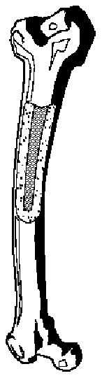 TOPICS OF STUDY – WEEK 10
 TOPICS OF STUDY – WEEK 13
 TOPICS OF STUDY – WEEK 14
 difficulty in completing this exam successfully.