Fibrin clot adhesion and root conditioning EVALUATION OF FIBRIN CLOT ADHESION AS A PRECURSOR FOR NEW ATTACHMENT FOLLOWING ROOT CONDITIONING ABSTRACT The objective of the present study was to evaluate the influence of various root conditioningmodalities on adsorption and adhesion of blood to scaled and planned root surfaces. This research workwas done in the Department of Periodontics, DAV Dental College, Yamunanagar, Haryana, India (Year2006-2009). The SEM analysis was done in …SEM & TEM centre, Department of Radiology, AIIMS,New Delhi (India)A total of 24 specimens were obtained from fresh extracted human teeth which were divided into4 groups comprising of 6 specimens in each group. The root conditioning groups included 3experimental – Citric acid, EDTA (Ethylene diamine tetra acetic acid), Tetracycline hydrochloride and1 PBS (Phosphate buffered saline) as control group. The root surfaces were planned and specimenblocks (4x4x1mm) were obtained. They were subject to various conditioning agents and then exposedto fresh blood which was allowed to clot. These specimens were then rinsed and subjected to SEManalysis.The results showed that Citric acid treated planned root specimens presented a dense blood cellsand fibrin attachment, while tetracycline treated showed moderate and EDTA treated showed scarceattachment of blood clot elements. In contrast, untreated planned dentin exhibited smear layer. Theadhesion of fibrin network and blood cell attachment was best following application of Citric acidfollowed by moderate attachment seen in Tetracycline.Key words: Root-conditioning, Blood, Fibrin, Smear INTRODUCTION
nated cementum. But such mechanical debridementand planning of teeth generate a smear layer. Root
The ultimate goal of periodontal therapy is the conditioning by topical application of acidic solutions
regeneration of the supporting tissues at the site has been demonstrated to remove not only this smear
previously exposed by periodontal disease.1 One of layer but also any remaining root surface contami-
major hindrance in inhibiting predictable regenera- nants. A number of agents have been proposed for the
tion appears to be the nature of the periodontitis demineralization including Phosphoric acid, EDTA,
affected root surface. Complex inflammatory, enzy- Citric acid. Tetracycline and Fibronectin.2 Citric acid
matic and other biological influences which accompany and tetracycline exert their etching action through a
periodontal disease produce physical or chemical alter- low pH (1 and 1.3 respectively) while EDTA chelates
ations which are particularly apparent in root cemen- divalent cations, such as Ca2+ at neutral pH (7.5).
Surface demineralization of dentin by these agents
The traditional treatment of such pathologically exposes the collagen matrix, there by providing a
altered root surfaces has relied on mechanical removal substrate that supports the chemotaxis, migrationof plaque and calculus, root bound toxins, and contami- and attachment of cells involved in wound healing
1 Senior Lecturer, Department of Periodontics, Dasmesh Institute of Research and Dental Sciences, Faridkot,
Correspondence: 2Dr Pardeep K Bansal, Department of Prosthodontics and Maxillofacial Prosthetics,
Dasmesh Institute of Research and Dental Sciences, Faridkot 151203 (Punjab), India, Mobile: 09814282284,
Fax: 01639251666, Email: [email protected]Pakistan Oral & Dental Journal Vol 30, No. 2 (December 2010) Fibrin clot adhesion and root conditioning
and formation of new connective tissue attach-
to obtain tetracycline HCl solution of pH 1.3 as
In vitro studies have indicated that cells attach
b) Citric Acid: 65 grams of anhydrous citric acid
better to etched than to non etched dentin surfaces,
crystals (Glaxo Smith Klein Co) were dissolved
regardless of choice of etching agent.6 The dentinal
in 100 ml of distilled water at room tempera-
tubules and intra – and peritubular collagen matrix
ture till the solution became saturated to ob-
exposed by these acids and chelating agents can sup-
tain citric acid at pH 1 which was checked using
port adhesion of fibrin clot.7 This is important as the
first requirement for successful regeneration rests
c) EDTA: 15% EDTA solution was prepared by
with clot adhesion to the root surface, possibly by
combining 25 ml of distilled water with 2.31 ml
of 5 N NaOH (Glaxo Smith Klein Co), and then
Various experimental studies have suggested that
adding 4.25 gm of the disodium salt of EDTA
periodontal regeneration is dependent upon the ad-
(Glaxo Smith Klein Co). This solution had a pH
sorption, uninterrupted adhesion and maturation of
the fibrin clot positioned between the gingival flap and
Each of the four groups analysed in this study
a periodontally compromised root.9 The blood elements contained 6 samples: 1) Immersed in citric acid solution
imposed must establish an attachment that endures 2) in tetracycline hydrochloride solution 3) in EDTA
the forces and remain stable until sufficient tensile solution 4) in PBS.
strength is achieved by the matured tooth gingivalinterface.7 This rapid adherence of blood clot to the root
Immersion was carried out for 5 minutes. After
surface may thus form a sufficient barrier to apical conditioning, three-5 minutes washes in PBS were
epithelial cell migration and lead to connective tissue carried out. After this, a healthy female donor was
attachment following periodontal regenerative sur- hematologically tested and a drop of her whole periph-
eral blood applied to external dentin surface on eachroot block. The blood was allowed to clot for 20 minutes
METHODOLOGY
in a humidifier chamber. Block were than rinsed three
times for 5 minutes in PBS. Washes and rinses werecarried out in small petri dish with gentle swirling
Twenty-four dentin blocks, approximately 4x4x1mm motion. All steps were carried out at room tempera-
in size were prepared from roots of freshly extracted ture.15
human teeth. The tooth included in the study werethose affected by periodontal disease, characterized by SEM analysis preparationbleeding on gentle probing, grade III mobility, radio-graphic evidence of proximal bone loss. The exclusion
Immediately after rinsing, the blocks were fixed in
criteria included the presence of caries, history of 1% formaldehyde in PBS for 15 minutes followed byscaling, root planning in the previous 6 months, teeth three 5 minute PBS rinses. After this the blocks werewith periapical infection or nonvital teeth and patients incubated for 10 minutes in 0.02 M glycine in PBS andwith history of systemic disease. The samples were rinsed again, as before. The samples were post-fixed inobtained after planning of root surfaces and stored in 2.5% glutaraldehyde in PBS for 30 minutes and rinsedPBS (phosphate buffered saline) pH 7.4 at 4°C again, as above. The samples were then dehydrateduntil use.
through a graded ethanol series: 25%, 50%, 75%, 95%
and 3 exchanges of 100%, all steps at room tempera-
ture. The teeth were kept for fifteen minutes in allgraded series and for half an hour in 100% acetone.11
a) Tetracycline HCl: 500 mg of tetracycline HCl
capsule (Hostacycline,™ Aventis Co) was dis-
The samples were subsequently dried overnight in
solved in 5 ml of sterile water i.e. distilled a dehydration jar. The dentine blocks were mounted onwater under continuous stirring for 10 minutes aluminium stubs with an adhesive tape with the labial
Pakistan Oral & Dental Journal Vol 30, No. 2 (December 2010) Fibrin clot adhesion and root conditioning
side of the roots facing the beam of the SEM and in sucha ways that the root were placed in the center of thestubs. They were vacuum coated with a thin layer ofcarbon, sputter coated with gold/palladium. The goldcoating was done to ensure a proper conducting surfaceto the non-conducting specimen. The coated specimenswere removed from the sputter coating unit and thenthe stubs were mounted on the specimen stage of theSEM unit. Observations were performed with a scan-ning electron microscope (Jeol JSM, Tokyo, Japan) at15.0 KV and observed on the computer screen fittedwith SEM. Photomicrographs of representative areaswere taken with a 35-mm camera.11
All specimens were examined and one photomicro-
graph obtained from a random area treated with bloodtissue at 2000x magnifications using scanning electronmicroscope operated at an accelerated voltage of 15KV. After the photomicrographs were obtained, theywere identified and analyzed through scores in order toverify the adhesion of blood components and to analyzethe morphological characteristics of root surface ob-tained after treating with various root conditioningagents. Using a single blind method, the photomicro-graphs obtained from samples that received bloodtissue were examined three times by an operator whowas previously trained. It was again calibrated withtwo other operators. Each sample received the scorethat prevailed among the three readings.15,16
Score 0: Absence of fibrin network and blood cells
Score 1: Scarce fibrin network and/or blood cells
Score 2: Moderate fibrin network and moderate quan-
Score 3: Dense fibrin network and trapped blood cells
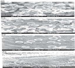
Statistical analysis was done with standard com- Fig 1: Photomicrographs of the root surfacesshowing
puter software. The non parametric Kruskal Wallis
a) dense red blood cells and fibrin attachment
aftertreatment with CA, b) moderate red blood
test (p<0.05) was employed to compare the rank of the
cells and fibrin attachment after treatment
evaluated groups. This procedure was followed by non
with TTC HCl, c) scarce red blood cells and
parametric Mann-Whitney U test when the Kruskal
fibrin attachment after treatment with EDTA,
Wallis test suggested a significant difference between
d) smear layer after treatment with PBS (Origi-
the groups (p<0.05). The Mann-Whitney U test (P<0.05)
Pakistan Oral & Dental Journal Vol 30, No. 2 (December 2010) Fibrin clot adhesion and root conditioning
Fig 2: Score distribution according to the different treatments performed
was calculated to determine differences between indi-
In group 2 i.e TTC HCl treated samples, four
samples showed moderate fibrin and blood cells attach-ment (score 2) while only two samples showed scarce
fibrin and blood cells attachment (score 1) (Fig 1b). Scanning electron microscopy analysis
In group 3 i.e EDTA treated samples, all the 6
sample showed scarce fibrin and blood cells attachment
In group 1 i.e CA treated samples, there was (score 1) (Fig 1c).
dense fibrin network and entrapped blood cells (score3) in five samples while one sample showed mo-
In group 4 i.e PBS treated samples (control), none
derate fibrin and blood cells attachment (score 2) of the 6 samples showed any attachment of fibrin or(Fig 1a).
Fig 3: Shows inter group comparison of adhesion of fibrin and blood cells between different groups using mean
Pakistan Oral & Dental Journal Vol 30, No. 2 (December 2010) Fibrin clot adhesion and root conditioning
TABLE 1: ALLOCATION AND COMPARISON OF ADHESION OF BLOOD COMPONENTS AND FIBRIN
Absence of Scarce fibrin Moderate Dense fibrin fibrin net- network and/ fibrin net- or blood cells and trapped blood cells (Score 1) blood cells blood cells (Score 0) (Score 2) (Score 3) Comparison specimens Statistical Results
score was highest in group 1, in the same way, group 2showed higher scores for blood components attach-
Considering groups as an independent variable, ment when compared to group 3 and 4.
the use of non parametric Kruskal Walli test showed a significant difference among the evaluated groups re- DISCUSSION garding blood components adhesion scores (p=0.000)
Root surface conditioning by topical application of
acidic solutions has been demonstrated to remove the
The comparison between the mean ranks of the smear layer and also any remaining root surface
groups (Mann-Whitney U test) showed a significant contaminants.1 It uncovers and widens the orifice ofdifferences between group 1 and group 2 (p=0.006), dentinal tubules12 with unmasking of the intra-andgroup 1 and group 3 (p=0.001), group 1 and group 4 peri-tubular dentin collagen matrix6,12 due to deminer-(p=0.001), group 2 and group 3 (p=0.019), group 2 and alization. This is indispensable for adhesion of fibringroup 4 (p=0.002), group 3 and group 4 (p=0.001) (Fig 3). clot.
When group 1 was compared with group 2, 3 and 4,
The clot adhesion appears vitally dependent on the
it was observed that the blood components adhesion formation of a resilient unit between the clot (fibrin
Pakistan Oral & Dental Journal Vol 30, No. 2 (December 2010) Fibrin clot adhesion and root conditioning
network) and the collagen fibers exposed at the root tion; 2) dentin conditioning to remove the instrumen-surface.7,15
tation surface smear layer; 3) formation of the fibrinclot. It is conceivable that those conditions that may
The present study was designed to evaluate the promote or adversely affect fibrin clot adhesion in this
extent of the fibrin clot adhesion on cleansed root model system may also produce similar effects in vivo.
The study sample consisted of 24 single rooted
Instrumentation of the human dentin blocks pro-
human teeth, affected by periodontitis with grade III duced a smear layer as has been shown in previousmobility extracted from patients with no systemic reports.22,23 Conditioning of the instrumented dentindisease.
with a saturated citric acid solution, tetracycline hy-drochloride solution or EDTA solution, but not PBS, at
Only single rooted teeth were selected for the least in part, removed the smear layer exposing den-
purpose of standardization. Teeth affected by caries tinal tubules and the intra and inter tubular collag-
were not included in the study as it could adversely enous matrix. This observation is in harmony with a
affect the root surface topography.17 Minimal instru- large number of reports evaluating the effect of acidic
mentation during extraction was considered to avoid and chelating agents on the ultrastructure of dentin
chipping off root structure.18,19 Teeth with immediate surfaces.6,7,11,23
past history of scaling and root planning procedureswere excluded from the study as these procedures may
Group 1 (Citric acid) exhibited vast three dimen-
alter the root surface. Teeth with attrition, abrasion sional array of dense inter connected fibrin strandsand erosion were excluded as they are shown to enmeshing the trapped blood cells (seen in 5 samples)produce secondary changes in the tooth structure like to moderate fibrin network and blood cells (seen in 1alteration in mineral composition and the formation of sample). sclerotic dentin.20
When citric acid was compared with other groups,
The obtained specimens were categorized into 4 the attachment of fibrin and blood cells was statistically
groups (one control and three experimental) consisting significant. The mean rank of citric acid was 9.17 whileof 6 specimens in each group. Phosphate buffered for TTC was 3.83, mean rank of citric acid was 9.50saline was used for temporary storage of the teeth. The while for EDTA was 3.50 and mean rank of citric acidsolution used during this short holding time should not was 9.50 while for PBS was 3.50 (Table 2). have affected the final surface characteristics.21
The possible explanation of the results seen in the
The root specimens were immersed in root condi- citric acid could be that saturated citric acid success-
tioning agents for five minutes. This is in accordance to fully removes the smear layer24,25 and provides greaterthe study done by Wen CR et al (1992)13 showing that
depth of demineralization as compared to TTC HCl as
wide opening of dentinal tubules and tufting of inter well as increased tubular diameter, as was seen intubular dentin fibrils was highest in immersion group citric acid conditioned root surfaces.12,21 All this in-as compared to cotton pellet placement or burnishing creased the wettability of dentin resulting in enhancedtechnique or those treated with camel hair brush. The attachment of the fibrin clot imposed on to the rootapplication of root conditioning agents resulted in root surfaces, as continuous adhesion of fibrin clot appearssurface demineralization exposing dentinal tubules dependent on the wettability of the substrate.7 It isand collagen of intra-and peritubular dentinal matrix11 observed that although citric acid is an anticoagulant,which leads to adhesion of fibrin clot.7
it has shown no adverse effects on early fibrin polymer-
provides a simulation of most of the critical steps
The results obtained in this study are in accordance
during the earliest healing events following periodon- to those obtained by Baker PJ et al (2000)11; Baker DL
tal tissue regeneration procedures. This method allows et al (2005)7 in their in vitro studies. Also, Polson AM
observations of the dentin surface directly following (1983)26 have shown the efficacy of citric acid condition-
various stages of simulation i.e 1) root instrumenta- ing to support maturation of fibrin clot into a new
Pakistan Oral & Dental Journal Vol 30, No. 2 (December 2010) Fibrin clot adhesion and root conditioning
connective tissue attachment using a non human
The root specimens treated with EDTA (Group III)
demonstrated scarce fibrin network and blood cells. When EDTA was compared to PBS, the attachment of
In contrast, animal studies by Nyman et al (1981), fibrin and red cells was found to be statistically signifi-
Gottlow et al (1984) and clinical trials by Stahl and cant. The mean rank of EDTA was 9.50 while for PBS
Froum (1977); Stahl et al (1983), Smith et al (1987) was 3.50 (Table 2).
utilized citric acid for root conditioning have failed toresult in new attachment.5 Speculative explanation for
It was observed that most samples in group III
these in consistent findings have included variations in inhibited blood element adsorption and adhesion to theanimal models, inconsistent flap adaptation, inadequate root surface. The possible explanation could be thatdemineralization of periodontitis affected root surface3 EDTA is a calcium chelator and may have inhibited orand repopulation of the root surface with inappropriate retarded coagulation events.I5,30,31 It was also observedcell types.27
that EDTA did not consistently produce a smear layerfree dentin surface.7
Group II (TTC HCl) exhibited the presence of
moderate fibrin network and moderate attachment of
In contrast, studies conducted by Blomlof and
blood cells (in 4 samples) to scarce fibrin network and Lindskog (1995)6 have shown that etching at neutralblood cells (in 2 samples).
pH with EDTA have shown to be equally, if not moreefficient compared to agents operating at low pH in
When tetracycline was compared with other groups exposing collagen fibres on dentin surfaces.
i.e EDTA and PBS, attachment of fibrin and red cellswas statistically significant. The mean rank of TTC was
The root specimens treated with PBS (group IV)
8.50 while for EDTA was 4.50, the mean ranks for TTC demonstrated no fibrin network or blood cell attach-was 9.50 while for PBS was 3.50 (Table 2).
ment. This could be due to presence of smear layer onroot specimens after treatment with phosphate buff-
The possible explanation of these results seen in ered saline. Specific dentin topographic features such
tetracycline hydrochloride could be that tetracycline as dentinal tubules, intra-and inter-tubular collagen
hydrochloride was successful in removing the smear fibrils were not readily discernible.7,16 The results
layer and exposed the opening of dentinal tubules with obtained in this group are in contrast to the study done
collagen matrix.1,28 In the present study TTC HCl fared by Leite FRM et al (2005)15 who showed that untreated
better than EDTA group as regards to fibrin network planed dentin presented the best results with blood
and blood cell attachment. It can be due to greater cells entrapped in a thick web of fibrin. This could be
demineralization potential of tetracycline than EDTA. due to the more repeated 5 min washings with PBS
According to Claffey et al 1987 more superficial dem- done after individual treatment with Formaldehyde,
ineralization obtained with tetracycline is responsible Glycine, Glutaraldehyde in this study which might
for a more favourable healing response. Several in vivo have washed the fibrin network 11 as compared to 5 min.
studies have demonstrated that tetracycline deminer- washings with PBS done only after Glutaraldehyde
alized dentin surfaces showed greater number of at- treatment by Leite FRM et al in their study.
tached cells24, greater connective tissue attachment28,29,reattachment and new cementum formation.28
In perspective, it appears reasonable to suggest
that in vitro protocols that sustain fibrin clot adhesion
In contrast, in a study done by Frantz B and Polson to dentin may support wound maturation into a connec-
A (1988)24 using a non human primate implantation tive tissue attachment in vivo. However, protocols that
model, it was shown that enhanced response to cell are less successful in vitro should not be expected to
attachment after demineralization by tetracycline did support fibrin clot adhesion in a clinical scenario.7
not result in a connective tissue attachment and tetra- cycline released from root surface altered the biological CONCLUSION
cascade of healing by altering PMN functions. AlsoDelazari FMC et al (1999)30 found fibrin network forma-
From this study it is concluded that the application
tion in situ was not improved by application of TTC-HCl. of citric acid and tetracycline on instrumented
Pakistan Oral & Dental Journal Vol 30, No. 2 (December 2010) Fibrin clot adhesion and root conditioning
periodontally diseased roots were better root condi- 16 Theodoro LH, Sampaio JEC, Haypek P, Bachmann L,tioning agents for the adhesion of the fibrin clot and
Zezell DM, Garcia VG. Effect of Er: Y AG and diode laserson the adhesion of blood components and on the morpho-
could prove to be a useful tool in the establishment of
logy of irradiated root surfaces. J Periodont Res 2006; 41:
a new connective tissue attachment in periodontal
17 Levespere JE, Yukana A. Root surface removal with diamond
coated ultrasonic instrument: An in-vitro and SEM study - JPeriodontol 1996; 67: 1281-87
REFERENCES
18 Ewen SJ, Gwinnett AJ. A scanning electron microscopic study
Trombelli L, Scabbia A, Zangari F, Griselli A, Wikesjo UME,
of teeth following periodontal instrumentation. J Perio-
Calura G. Effect of tetracycline HCI on periodontally affected
human root surfaces. J Periodontol1995; 66: 685-91
19 Khosravi M, Bahrami ZS, Atabaki MSJ, Shokrgozar MA,
Aravind B, Koshy C, Bhat GS, Bhat KM. Root conditioning -
Shokri F. Comparative effectiveness of hand and ultrasonic
instrumentation in root surface planing in vitro. J ClinPeriodontol2004; 31: 160-65
Hanes PJ, Polson AM. Cell and fiber attachment to deminer-alized cementum from normal root surfaces. J Periodontol
20 Ashwini S, Mehta DS. Comparative evaluation of surface
alterations on periodontally diseased root subsequent to theapplication of phosphoric acid, minocycline HCI and combina-
Hanes PJ, Polson AM, Frederick GT. Initial wound healing
tion of citric acid and tetracycline HCI- A scanning electron
attachments to demineralized dentin. J Periodontol 1988; 59:
microscopic study. JIDA 2001; 72:42-46
21 Lafferty TA, Gher ME, Gray JL. Comparative SEM study on
Polson AM, Hanes PJ. Cell and fiber responses to cementum
the effect of acid etching with tetracycline HCI or citric acid on
from periodontitis - affected root surfaces after citric acid
instrumented periodontally- involved human root surfaces. J
treatment. J Clin Periodontol 1989; 16: 489-97
Blomlof J, Lindskog S. Root surface texture and early cell and
22 Jones SJ, Lozdon J, Boyde A. Tooth surfaces treated In situ
tissue colonization after different etching modalities. Eur J
with periodontal instruments - Scanning electron microscopic
studies. Brit Dent J 1972; 132: 57-64
Baker DL, Stanley Pavlow SA, Wikesjo UME. Fibrin clot
23 Polson AM, Frederick GT, Ladenheim S, Hanes PJ. The
adhesion to dentin conditioned with protein constructs: an in
production of a root surface smear layer by instrumenta-
vitro proof-of-principle study. J Clin Periodontol2005; 32:
tion and its removal by citric acid. J Periodontol1984; 55:
Wikesjo UME, Claffey N, Egelberg J. Periodontal repair in
24 Frantz B, Polson A. Tissue interactions with dentin specimens
dogs - Effect of heparin treatment of the root surface. J Clin
after demineralization using tetracycline. J Periodontol1988;
Wikesjo UME, Selvig KA. Periodontal wound healing and
25 Hanes P, Polson A, Frederick T. Citric acid treatment of
regeneration. Periodontol2000. 1999; 19: 21-39
periodontitis - affected cementum. J Clin Periodontol1991;
10 Wikesjo UME, Crigger M, Nilveus R, Selvig KA. Early healing
events at the dentin connective tissue interface. Light and
26 Polson AM, Proye MP. Fibrin linkage: A precursor for new
transmission electron microscopy observations. J Periodontol
attachment. J Periodontol1983; 54: 141-47
27 Polson AM, Hanes PJ. Cell and fiber attachment to deminer-
11 Baker PJ, Rotch HA, Trombelli L, Wikesjo UME. An invitro
alized dentin.JClinPeriodontoI1987; 14: 357-65
screening model to evaluate root conditioning protocols forperiodontal regenerative procedures. J Periodontol2000; 71:
28 Alger FA, Solt CW, Vuddhakanok S, Miles K. The histologic
evaluation of new attachment in periodontally diseased hu-man roots treated with tetracycline - hydrochloride and
12 Labahn R, Fahrenbach WH, Clark SM, Lie T, Adams DF. Root
fibronectin. J Periodontol 1990; 61: 447-55
dentin morphology after different modes of citric acid andtetracycline hydrochloride conditioning. J Periodontol1992;
29 Wikesjo UME, Claffay N, Christersson LA, Franzetti LC,
Genco RJ, Terranova VP. Repair of periodontal furcationdefects in beagle dogs following reconstructive surgery in-
13 Wen CR, Caffesse RG, Morrison EC, Nasjleti CE, Parikh UK.
cluding root surface demineralization with tetracycline hy-
In vitro effects of citric acid application techniques on dentin
drochloride and topical fibronectin application. J Clin
surfaces. J Periodontol1992; 63: 883-89
14 Lasho DJ, O’Leary TJ, Kafrawy AH. A scanning electron
30 Delazari FMC, Gerlach RF, Joly JC, Lima AFM. Scanning
microscope study of the effects of various agents on instru-
electron microscopy study of the effect of tetracycline Hel on
mented periodontally involved root surfaces. J Periodontol
smear layer removal and fibrin network formation. Braz Dent
15 Leite FRM, Moreira CS, Theodoro LH, Sampaio JEC. Blood
31 B1omlof J, Lindskog S. Periodontal tissue-vitality after
cell attachment to root surfaces treated with EDTA gel. Braz.
different etching modalities. J Clin Periodontol1995; 22:
Pakistan Oral & Dental Journal Vol 30, No. 2 (December 2010)
LIDOKAIN BIZTONSÁGI ADATLAP 1907/2006 EK rendelet (REACH) és 1272/2008 EK rendelet (CLP-GHS) szerint 1. AZ ANYAG/KEVERÉK ÉS A VÁLLALAT/VÁLLALKOZÁS AZONOSTÁSA 1.1. Term ékazonosító A z anyagnak egyelõre nincs regisztrációs száma, mert a regisztráció késõbbi idõpontban várható, vagy az éves gyártott mennyiség alapján regisztráció nem szükséges, vagy a R
Opening: The regular meeting of the Pinchbeck Elementary School PTA Board was held on May 8, 2007 in the school library. The meeting was cal ed to order at 5:30pm by the President, Amy Farina. Approval of Minutes: The minutes from the April 24, 2007 meeting were approved. Vice-President – Michel e West reported that there wil be a Mad Science program for the 1st grade in June. The Internati
 Fibrin clot adhesion and root conditioning