Die Struktur von Tadalafil erlaubt eine selektive Bindung an die Bindungsstelle der PDE5 und minimiert gleichzeitig die Interaktion mit PDE6, was visuelle Nebenwirkungen einschränkt. Seine Verteilung im Organismus erfolgt breit, wobei das Verteilungsvolumen etwa 63 Liter beträgt. Über 90 % des Wirkstoffs sind an Plasmaproteine gebunden. Die Wirkung bleibt unabhängig von der Nahrungsaufnahme konstant. Der Abbauweg über CYP3A4 kann durch Hemmer wie Ritonavir oder Ketoconazol verlangsamt werden, was die Plasmakonzentrationen deutlich erhöht. In diesem Kontext wird cialis 20mg preis häufig in Bezug auf pharmakokinetische Wechselwirkungen erwähnt.
Doi:10.1016/j.jcrs.2005.10.030
J CATARACT REFRACT SURG - VOL 31, OCTOBER 2005
Management of infectious keratitis following laser
Eric D. Donnenfeld, MD, Terry Kim, MD, Edward J. Holland, MD, Dimitri T. Azar, MD, F. Rick Palmon, MD,
Jonathan B. Rubenstein, MD, Sheraz Daya, MD, Sonia H. Yoo, MD
Laser in situ keratomileusis (LASIK) is the most commonly
not diagnosed on initial presentation. Nine patients re-
performed refractive surgical procedure for the correction
quired flap excision, and 1 flap sloughed spontaneously.
of ametropia. The advantages of LASIK include rapid visual
One case required enucleation, and 10 required penetrating
rehabilitation, decreased stromal scarring, less irregular
keratoplasty for visual rehabilitation.
astigmatism, minimal regression, less postoperative pain,
In most cases, it is difficult to determine the origin of
and the ability to treat a greater range of refractive disor-
the infection. A recent ASCRS survey of infectious keratitis
ders.Unlike surface-ablation procedures, LASIK pre-
following photorefractive keratectomy (PRK) and LASIK
serves the integrity of Bowman’s membrane and the
for the year 2004 (Donnenfeld, ASCRS 2005) revealed a
overlying epithelium, thus decreasing the risk for microbial
significant decrease in atypical mycobacteria, with only
keratitis. However, microbial keratitis following LASIK has
2 cases reported. Prophylaxis with a fourth-generation
become an increasingly recognized, sight-threatening com-
fluoroquinolone was not done in either case. Sixty-one per-
cent of cases reported in this survey were due to Staphylo-
The incidence of infectious keratitis following LASIK is
coccus bacteria. Forty-eight infections were reported by
difficult to estimate and can vary widely depending on the
46 surgeons who had performed an estimated 102 300 pro-
source of the information. One large retrospective study in-
cedures; an incidence of 1 infection for every 2131 proce-
vestigating the complications associated with LASIK found
dures performed during the year 2004.
an incidence of 2 infections in 1062 ; a similar study
A review of the published reports of LASIK-associated
found an incidence of 1 infection in 1019 eyes.A more re-
microbial keratitis in the peer-reviewed literature reveals
cent case series of LASIK-associated infections encountered
over 100 cases with a striking preponderance of atypical
at a single institution reports an estimated incidence be-
mycobacterial (47%) and staphylococcal (19%) species.
tween 1:1000 and Based on a comprehensive re-
Another interesting yet concerning observation regarding
view and analysis of the literature on infections following
these atypical mycobacterial LASIK infections involves
LASIK, Chang et state that the incidence of infection
their ability to occur in clusters or epidemics. Separate clus-
after LASIK can vary widely (0% to 1.5%). In a survey by
ters of atypical mycobacterial infections following LASIK
the American Society of Cataract and Refractive Surgery
have been published in the peer-reviewed literature. Chan-
(ASCRthe incidence reported by LASIK surgeons
dra et al.report a series of 7 eyes in 4 patients, all of whom
who had experienced an infectious keratitis was 1 in
had hyperopic LASIK at the same surgery center by the
2919 cases performed during the year 2001. In this study
same surgeon on the same day. The causative organism, My-
of 116 cases, 76 presented the first week after surgery, 7
cobacterium chelonae, presumably originated from a contact
during the second week, 17 between the second week
lens that was used intraoperatively to mask a portion of the
and the fourth week, and 16 after 1 month. The most com-
laser’s ablation. Another cluster is reported by Fulcher and
mon organisms cultured were atypical mycobacteria (33 of
coauthin 7 eyes of 7 patients; Mycobacterium szulgai
116 cases, 28%) and staphylococci (23 of 116 cases, 20%)
was traced back to the ice that was used to chill BSS on
species. In 47 of the 116 cases, infectious keratitis was
the surgical field. Freitas et al.report a cluster of infectionsin 11 eyes of 10 patients; M chelonae was found in the por-table steaming unit used to clean the microkeratome. An-other large cluster that has not been published but has
Accepted for publication August 5, 2005.
been investigated by the Centers for Disease Control and
Authors are members of the American Society of Cataract and
Prevention (CDCP) occurred in a surgery center in Georgia
Refractive Surgery Cornea Clinical Committee.
and involved 24 patients presumed to be infected with
No author has a proprietary or financial interest in any material or
Mycobacterium gordonaeKarp et al.report a series of
sporadic cases of atypical mycobacteria. Based on their
SPECIAL REPORTS: MANAGEMENT OF POST-LASIK INFECTIOUS KERATITIS
findings, the CDCP concluded that LASIK-associated kera-titis from atypical mycobacteria may be more common thanpreviously thought and also suggested that LASIK could bea risk factor for the development of atypical mycobacterialkeratitis.
Infectious keratitis is a potentially devastating compli-
cation of LASIK. In addition to the ASCRS survey findingsregarding the morbidity of these infections,the series ofclustered atypical mycobacterial infections (25 eyes of 21patients) shows that 4 patients experienced bilateral infec-tions, almost 50% of the affected eyes required flap ampu-tation, and all patients required aggressive topical and oralantimicrobial therapy for a 2- to 3-month period.
The organisms encountered in infectious keratitis fol-
lowing LASIK can be unusual, difficult to predict, andwill often not respond to empiric therapy with older-gener-ation topical fluoroquinolone antibiotic agents. For this
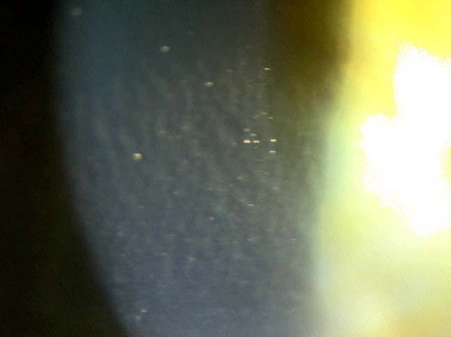
Figure 1. Clinical photograph of DLK showing diffuse intralamellar
reason, we highly recommend lifting the flap and taking
corneal scrapings for appropriate stains and cultures ifany suspicious infiltrate appears following LASIK. The re-
PREVENTION OF INFECTIOUS KERATITIS FOLLOWING LASIK
sults of these stains and cultures can be helpful in guidingantimicrobial therapy. A high degree of suspicion coupled
Several steps may help prevent infectious keratitis fol-
with a rapid diagnosis and appropriate therapy can result
lowing LASIK. Preoperatively, the lids and lacrimal appara-
in eradication of the infection and visual recovery. We rec-
tus of all patients considering refractive surgery should be
ommend that any focal infiltrate following LASIK should be
thoroughly examined. Treatment of infectious lid disease
considered infectious, and we discourage the practice of
with hot compresses and an antibiotic ointment applied 3
empirical antibiotic treatment without culturing.
times a day to the lid margin may help reduce the risk for
Diffuse lamellar keratitis (DLK) is a sterile inflamma-
bacterial keratitis. Proper sterilization techniques can pre-
tion of the lamellar interface following LASIK and is associ-
vent the use of contaminated instruments. A minority of
ated with epithelial abrasions and trauma. It traditionally
clinicians recommend performing monocular surgery or
occurs within the first few days after LASIK unless there
using separate instruments when performing bilateral sur-
is postoperative ocular trauma.Therapy is high-dose
gery,although this is not the practice of the members of
topical corticosteroids; in severe cases, oral corticosteroids
the ASCRS Cornea Clinical Committee. Some clinicians
and interface irrigation may be necessary
recommend the use of sterile drapes, gowns, gloves, and
Infectious keratitis following LASIK often presents with
inflammation in the corneal interface, which can mimicDLK. Because of this, many cases are typically treatedwith frequent topical corticosteroid therapy that can cloudthe clinical picture with transient improvement in the in-flammation. However, unlike DLK, the inflammation asso-ciated with LASIK-associated infections usually persistsdespite topical corticosteroids, and the underlying infec-tions can potentially worsen with corticosteroid tapering. The appearance of an interface inflammation more than 1week after LASIK should be presumed to be of an infectiousetiology until proven otherwise. Diffuse lamellar keratitischaracteristically has a diffuse appearance (asthe name suggests, while infectious keratitis has a focalarea of infiltration surrounded by diffuse inflammation(or even focal inflammation limited to the areaof the infiltrate. Any focal infiltrate surrounded by inflam-mation should be presumed infectious until proven
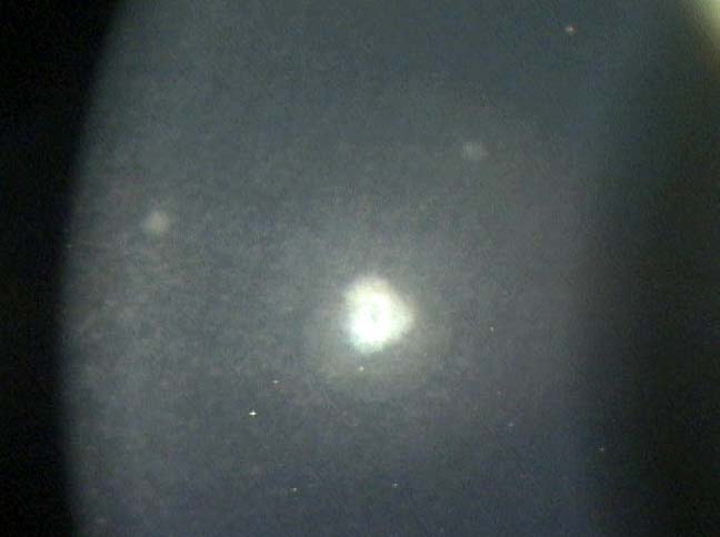
Figure 2. Clinical photograph of infectious keratitis following LASIK with
a focal infiltrate surrounded by diffuse inflammation.
J CATARACT REFRACT SURG - VOL 31, OCTOBER 2005
SPECIAL REPORTS: MANAGEMENT OF POST-LASIK INFECTIOUS KERATITIS
masks by the treating physician and assisting technician. A
0.5% given in a loading dose every 5 minutes for 3 doses
povidone–iodine solution (Betadine 10%) lid prep before
and then every 30 minutes, alternating with an antimicro-
cataract surgery has been shown to reduce the incidence
bial that is rapidly bacteriocidal and has increased activity
of endophthalmitis postoperatively and is recommended
against gram-positive organisms, such as fortified cefazolin
by many clinicians when performing LASIK.Finally, sev-
50 mg/mL every 30 minutes. In patients who work in a hos-
eral epidemics of atypical mycobacteria have been associ-
pital environment, there is an added risk for methicillin-re-
ated with the use of nonsterile water to clean instruments
sistant Staphylococcus aureus (MRSA). In these patients,
or the use of ice during LASIK.All fluids applied to
we recommend the substitution of fortified vancomycin
the eye before, during, and after LASIK should be sterile.
50 mg/mL for cefazolin every 30 minutes to provide moreeffective therapy against MRSA In addition,we advocate the use of oral doxycycline 100 mg twicea day to inhibit collagenase production and also discontin-
TREATMENT OF INFECTIOUS KERATITIS FOLLOWING LASIK
We divide infectious keratitis following LASIK into
For delayed-onset keratitis, which is commonly due to
early onset (occurring within the first 2 weeks of surgery)
atypical mycobacteria, nocardia, and fungi, we recommend
and late onset (occurring 2 weeks to 3 months after sur-
beginning therapy with amikacin 35 mg/mL every 30 min-
gery). The organisms seen in early-onset infectious keratitis
utes, alternating with a fourth-generation fluoroquinolone
are common bacterial pathogens such as staphylococcal
(gatifloxacin 0.3% or moxifloxacin 0.5%) every 30 min-
and streptococcal species. Gram-negative organisms are
utes, starting oral doxycycline 100 mg twice a day, and dis-
rare. The organisms seen in late-onset infectious keratitis
continuing corticosteroids (This treatment will
are usually opportunistic such as fungi, nocardia, and atyp-
not affect fungal infections; therefore, treatment in all cases
ical mycobacteria. The literature review of LASIK-associated
of infectious keratitis should be modified based on culture
infections by Chang and coauthorssupports this classifi-
and scraping results and clinical response to therapy.
cation of infection. Based on their study, gram-positive
In conclusion, infectious keratitis is a potentially dev-
organisms were more likely to present within 7 days of sur-
astating complication following LASIK. Culture results re-
gery (P Z.001) while mycobacterial infections were more
veal opportunistic infections and gram-positive bacteria as
likely to present 10 or more days after surgery (P!.001).
the most common organisms. Infectious keratitis may pres-
Since the organisms responsible for infectious keratitis
ent as late as months after LASIK, and its frequent misdiag-
following LASIK will often not respond to empiric therapy,
nosis at initial presentation may result in significant vision
we recommend lifting the flap, scraping and culturing sus-
loss. We do not recommend empiric therapy as most organ-
picious cases, and selecting appropriate culture media in-
isms are opportunistic and do not respond to conventional
cluding blood agar, chocolate agar, Sabouraud’s agar, and
therapy. A high degree of suspicion with flap elevation and
thioglycolate broth. For infectious keratitis after 2 weeks,
culturing should be performed in all eyes suspected of
we recommend a growth media for atypical mycobacteria
having an infectious infiltrate(s) following LASIK.
such as Lowenstein-Jensen or Middlebrook 7H-9 agar in
We hope the information contained in this report will
addition to the other culture media. If these special media
help LASIK surgeons assess their respective approaches to
are unavailable, we recommend using blood agar as atypi-
the management of post-LASIK infectious keratitis. The
cal mycobacteria grow quite well on these plates. At thetime of culture, we also recommend scraping the infiltrate
Elevate flap
and performing a Gram stain, Gomori-methenamine silverstain, and Ziehl-Neelsen stain to rule out unusual patho-
Culture and scrape
gens such as nocardia, atypical mycobacteria, and fungi. Onset 2 Weeks or Less
In cases in which cultures are negative and the infectioncontinues to worsen, a corneal biopsy or polymerase chain
gatifloxacin 0.3 or moxifloxacin 0.5 alternating with cefazolin 50 mg/mL every 30 minutes
For the treatment of rapid-onset and delayed-onset in-
If patient is exposed to hospital environment, substitute vancomycin 50 mg/mL
fectious keratitis, the recommendation is to elevate the flap
for cefazolin
and culture. Irrigation of the flap interface with an appro-
Onset 2 Weeks or More
priate antibiotic solution (fortified vancomycin 50 mg/mLfor rapid-onset keratitis and fortified amikacin 35 mg/mL
gatifloxacin 0.3 or moxifloxacin 0.5 alternating with
for delayed-onset keratitis) may be helpful. For rapid-onset
amikacin 35 mg/mL every 30 minutes
keratitis, we recommend a fourth-generation topical fluo-roquinolone such as gatifloxacin 0.3% or moxifloxacin
Figure 3. Treatment of infectious keratitis following LASIK.
J CATARACT REFRACT SURG - VOL 31, OCTOBER 2005
SPECIAL REPORTS: MANAGEMENT OF POST-LASIK INFECTIOUS KERATITIS
goal is to standardize treatment, minimize visual loss, and
10. Chang MA, Jain S, Azar DT. Infections following laser in situ keratomi-
leusis: an integration of the published literature. Surv Ophthalmol2004; 49:269–280
11. Solomon R, Donnenfeld ED, Azar DT, et al. Infectious keratitis after la-
ser in situ keratomileusis: results of an ASCRS survey. J Cataract RefractSurg 2003; 29:2001–2006
1. Hersh PS, Brint SF, Maloney RK, et al. Photorefractive keratectomy ver-
12. Chandra NS, Torres MF, Winthrop KL, et al. Cluster of Mycobacterium
sus laser in situ keratomileusis for moderate to high myopia; a ran-
chelonae keratitis cases following laser in-situ keratomileusis. Am J
domized prospective study. Ophthalmology 1998; 105:1512–1522;
13. Fulcher SFA, Fader RC, Rosa RH, Holmes GP. Delayed-onset mycobac-
2. Pallikaris IG, Papatzanaki ME, Stathi EZ, et al. Laser in situ keratomileu-
terial keratitis after LASIK. Cornea 2002; 21:546–554
14. Freitas D, Alvarenga L, Sampaio J, et al. An outbreak of Mycobacterium
3. Pe´rez-Santonja JJ, Bellot J, Claramonte P, et al. Laser in situ keratomi-
chelonae infection after LASIK. Ophthalmology 2003; 110:276–285
leusis to correct high myopia. J Cataract Refract Surg 1997; 23:372–
15. Winthrop KL, Steinberg EB, Holmes G, et al. Epidemic and sporadic
cases of nontuberculous mycobacterial keratitis associated with laser
4. Helmy SA, Salah A, Badawy TT, Sidky AN. Photorefractive keratectomy
in situ keratomileusis. Am J Ophthalmol 2003; 135:223–224
and laser in situ keratomileusis for myopia between 6.00 and 10.00 di-
16. Bu¨hren J, Kohnen T. Corneal wound healing after laser in situ kerato-
opters. J Refract Surg 1996; 12:417–421
mileusis flap lift and epithelial abrasion. J Cataract Refract Surg 2003;
5. Salah T, Waring GO III, El-Maghraby A, et al. Excimer laser in-situ ker-
atomileusis (LASIK) under a corneal flap for myopia of 2 to 20 D. Trans
17. Stulting RD, Randleman JB, Couser JM, Thompson KP. The epidemiol-
Am Ophthalmol Soc 1995; 93:163–183; discussion 184–190
ogy of diffuse lamellar keratitis. Cornea 2004; 23:680–688
6. Azar DT, Farah SG. Laser in situ keratomileusis versus photorefractive
18. Hoffman RS, Fine IH, Packer M. Incidence and outcomes of LASIK with
keratectomy; an update on indications and safety [guest editorial].
diffuse lamellar keratitis treated with topical and oral corticosteroids.
J Cataract Refract Surg 2003; 29:451–456
7. Stulting RD, Carr JD, Thompson KP, et al. Complications of laser in situ
19. Kohnen T. Infections after corneal refractive surgery: can we do bet-
keratomileusis for the correction of myopia. Ophthalmology 1999;
ter? (editorial) J Cataract Refract Surg 2002; 28:569–570
20. Speaker MG, Menikoff JA. Prophylaxis of endophthalmitis with topical
8. Lin RT, Maloney RK. Flap complications associated with lamellar re-
povidone-iodine. Ophthalmology 1991; 98:1769–1775
fractive surgery. Am J Ophthalmol 1999; 127:129–136
21. Kohnen T, Scho¨pfer D, Bu¨hren J, Hunfeld KP. Flapamputation bei My-
9. Karp CL, Tuli SS, Yoo SH, et al. Infectious keratitis after LASIK. Ophthal-
cobacterium chelonae-Keratitis nach Laser-in-situ-Keratomileusis. Klin
J CATARACT REFRACT SURG - VOL 31, OCTOBER 2005
l'alimentation et des produits los Productos de de consommation Certificate Gesundheitsbescheinigung Certificat ANIMAL HEALTH CERTIFICATE (REQUIREMENTS) FOR CAMELIDS TO BE EXPORTED TO JAPAN FROM THE NETHERLANDS premises of origin of the exported : camelids Date starting embarkation Paratuberculosis: delayed type hypersensitivity test using Johnin* or Avian and Fecal culture test* or
Physician Assessment Form (All Visits) Acne Stain Score (pigmentary changes from acne) Canadian Acne Epidemiological Survey B10. Face - Acne Stain Score. (Select ONE best response) ¡ Clear ¡ Almost clear ¡ Mild ¡ Moderate B11. Chest - Acne Stain Score. (Select ONE best response) ¡ Clear ¡ Almost clear ¡ Mild ¡ Moderate B12. Back - Acne Stain Score. (Select ONE best response)
 SPECIAL REPORTS: MANAGEMENT OF POST-LASIK INFECTIOUS KERATITIS
findings, the CDCP concluded that LASIK-associated kera-titis from atypical mycobacteria may be more common thanpreviously thought and also suggested that LASIK could bea risk factor for the development of atypical mycobacterialkeratitis.
SPECIAL REPORTS: MANAGEMENT OF POST-LASIK INFECTIOUS KERATITIS
findings, the CDCP concluded that LASIK-associated kera-titis from atypical mycobacteria may be more common thanpreviously thought and also suggested that LASIK could bea risk factor for the development of atypical mycobacterialkeratitis.