Die Struktur von Tadalafil erlaubt eine selektive Bindung an die Bindungsstelle der PDE5 und minimiert gleichzeitig die Interaktion mit PDE6, was visuelle Nebenwirkungen einschränkt. Seine Verteilung im Organismus erfolgt breit, wobei das Verteilungsvolumen etwa 63 Liter beträgt. Über 90 % des Wirkstoffs sind an Plasmaproteine gebunden. Die Wirkung bleibt unabhängig von der Nahrungsaufnahme konstant. Der Abbauweg über CYP3A4 kann durch Hemmer wie Ritonavir oder Ketoconazol verlangsamt werden, was die Plasmakonzentrationen deutlich erhöht. In diesem Kontext wird cialis 20mg preis häufig in Bezug auf pharmakokinetische Wechselwirkungen erwähnt.
The structure and function of macromolecules
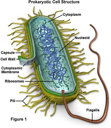
Julia Hughes HNC/D Animal Studies Module: Microbiology Microbiology: Practical Competence Introduction Infectious diseases in animals are caused by the invasion of tissues by bacteria, especially the epithelium, by microorganisms. This invasion have many effects which can be detrimental to the animals health, let alone be passed on to other animals through physical contact, aerosol, contamination of water or by being transmitted through a parasite such as the common flea. Prevention by vaccination, good hygiene, quarantine or culling is the best way of reducing and eliminating bacterial infections however it may be necessary to treat the infection with antibiotic and antibacterial drugs. These drugs help alleviate the symptoms together with the use of anti-inflammatory or analgesic drugs, rest and good nutrition. Viruses and bacteria are prokaryotes, simple structures that lacking well-defined nuclei and membrane-bound organelles and their own metabolism (see figure 1 below). They are all obligate parasites on plants or animal cells, hijacking the cells metabolism in order to produce more of the virus that continue to reproduce until the cell ruptures. Julia Hughes HNC/D Animal Studies Module: Microbiology
There are several different ways to test for bacteria in a laboratory format. Some culture types of which include:-
The growth rate will vary according to condition in temperature. A rise of 10% will double the growth rate and E-coli doubles every 20 minutes at body temperature.
Culture Tests with Micrococcus Luteus Micrococcus Luteus is gram positive, generally a non-pathogenic bacteria (but may be opportunistic in immunosuppressed individuals) and mainly considered a contaminant. It primarily habitats the normal flora of the mammalian skin, human mouth, mucosae, oropharynx and upper respiratory tract but can also be found in dust, air, water and soil. Results of Laboratory Testing Several experiments were performed using this bacteria as follows. In these experiments a nutrient agar or a meat extract, have been used. Preparation of a Petri Dish
1. The nutrient agar is warmed up and kept in a warm water bath, preferably at
body temperature to keep the culture from solidifying.
2. An empty sterile Petri Dish was opened slightly and quickly had the nutrient
agar poured into it in order to cover the bottom and fill the dish to approximately one third.
3. The dish is labeled with initials, date and contents of Nutrient Agar. 4. The nutrient is then left to set and can be used for experimenting.
Contamination of a Petri Dish
1. The lid of a pre-prepared Petri Dish was removed and left open to the room air
2. The lid was then replaced and the dish was stored at room temperature
On examination the following day, it was found that within the 10 minute exposure to air, the nutrient agar had become contaminated. This result confirms the fact that there is air-borne but non-harmful bacteria in normal air that is breathed every day.
Julia Hughes HNC/D Animal Studies Module: Microbiology A Streak Test
1. Prepare the area first by cleaning down the surfaces with 100% alcohol
2. Put a Bunsen burner on full flame to pull up the air around it and kill off any
3. Sterilise a loop in a high flame. 4. Take the lid off a slope agar with the bacteria Micrococcus Luteus, flame the
top of the bottle and then quench the heat of the loop first in the agar.
5. Scrap a tiny amount of bacteria from the slope, put the lid back on and sweep
the sample lightly three times across the top, three times down the side and then three times along the bottom of the agar in the Petri dish, keeping the lid as low down as possible to avoid any unnecessary contamination.
6. Reposition the lid of the Petri dish. 7. Reflame the loop to kill off any remaining bacteria. 8. The Petri dish is then turned upside down for labeling with initials, date and
sample name of M Luteus Streak and placed in a warm area of 37oC overnight.
The result of this test showed viable cells of bacteria growing in all three of the streaked areas (see photograph).
A Lawn Test
1. Prepare the area first by cleaning down the surfaces with 100% alcohol
2. Put a Bunsen burner on full flame to pull up the air around it and kill off any
3. Sterilise a loop in a high flame. 4. Take the lid off a slope agar with the bacteria Micrococcus Luteus, flame the
top of the bottle and then quench the heat of the loop first in the agar.
5. Scrap a tiny amount of bacteria from the slope, put the lid back on and drop
the sample gently onto the agar in the Petri dish. Reflame the loop to kill off any remaining bacteria.
6. Take a swab out of its packet and spread the bacteria evenly all over the agar,
keeping the lid as low down as possible to avoid any unnecessary contamination.
7. Reposition the lid of the Petri dish. 8. Reflame the loop to kill off any remaining bacteria. 9. The Petri dish is then turned upside down for labeling with initials, date and
sample name of M Luteus Lawn and placed in a warm area of 37oC overnight.
The result of this test showed viable cells of bacteria growing all over the agar (see photograph).
Julia Hughes HNC/D Animal Studies Module: Microbiology An Antibiotic Test
1. Prepare the area first by cleaning down the surfaces with 100% alcohol
2. Put a Bunsen burner on full flame to pull up the air around it and kill off any
3. Put some alcohol in a beaker. 4. Sterilise a loop in a high flame. 5. Take the lid off a slope agar with the bacteria Micrococcus Luteus and quench
6. Scrap a tiny amount of bacteria from the slope, put the lid back on and drop
the sample gently onto the agar in the Petri dish. Reflame the loop to kill off any remaining bacteria.
7. Take a swab out of its packet and spread the bacteria evenly all over the agar,
keeping the lid as low down as possible to avoid any unnecessary contamination.
8. Reposition the lid of the Petri dish. 9. Reflame the loop to kill off any remaining bacteria. 10. Put some tweezers in the beaker with the alcohol. 11. Pick up an antibiotic disc with the tweezers and lay it on the agar lawn. 12. The Petri dish is then turned upside down for labeling with initials, date and
sample name of M Luteus Antibiotic and placed in a warm area of 37oC overnight.
The result of this test was inclusive but should have shown which of the antibiotic discs had the greater affect of ensure that the bacteria did not grow. Where the antibiotic had no effect whatsoever, large growths of bacteria would have grown around and under that particular disc. Another test was performed and the results are shown in the photographs attached. Working from the top disc results show as follows:-
“FC” - Fusidic Acid – some intermittent clearing of bacteria
“OX” – Oxacillin – no bacteria cleared
“NO” – Novobiocin – bacteria very noticeable around the disc “PG” – Penicillin G – no change with bacteria growth steady
“S” – Streptomycin – virtually total clearance of bacteria “T” – Tetracycline – localize intense clearing of bacteria
“C” – Chloramphenicol – a wide clearing of bacteria
“E” – Erythromycin - a large clearing of bacteria
These results shows that while Fusidic Acid, Tetracycline and Chloramphenicol have an effect on Micrococcus Luteus, Streptomycin and Erythromycin are the most efficient in their anti-bacterial qualities.
Julia Hughes HNC/D Animal Studies Module: Microbiology References
GENDER ISSUES IN INTERNATIONAL TRADE by Marina Fe B. Durano 1. Introduction Trade policies have different consequences on women and men because women and men differ in their economic and social status. Women and men respond differently to economic and trade policies because they have different sets of private resources and levels of access to public ones. Status and control over resour
DOCUMENTOS PROCEDENTES DEL ARCHIVO HISTÓRICO MUNICIPAL DE ÚBEDA RELACIONADOS CON ASPECTOS DE LA VIDAD COTIDIANA DE LA CIUDAD DURANTE EL SIGLO XVII DOCUMENTO Nº 1 - Archivo Histórico Municipal de Úbeda. - Fondo de Protocolos Notariales. - Escribano: Francisco de Biedma. - Legajo nº 626. - El documento aparece en la contraportada. Memorial de medicinas administradas a un enfermo
 Julia Hughes